
 Products
Products
Products








On December 12, 2024, the research team led by Jia Jing from the School of Chemistry and Chemical Engineering, Nanjing University, published findings in Small. Supported by the Nexcope NCF Confocal Microscope, this study constructed cancer‑cell‑membrane‑camouflaged nanoprobe TCC@M based on Ti₃C₂ MXene. It integrates a photo‑dynamically activated CRISPR‑Cas9 system for targeted delivery and spatiotemporally controllable gene regulation. Combined with the photothermal properties of MXene, enhanced photothermal therapy is realized, while multi‑modal imaging (fluorescence / photoacoustic / thermal) is incorporated to guide treatment. This technology addresses clinical bottlenecks of conventional combined cancer therapy, including poor synergy, single spatiotemporal control, lack of real‑time monitoring, and limited tissue penetration of light‑controlled gene regulation. It achieves efficient cancer therapy under mild temperatures with favorable targeting, safety and precision, offering an innovative synergistic strategy for precise cancer diagnosis and treatment.
Research Background
Owing to the complexity of the tumor microenvironment, traditional cocktail‑style combination therapy delivers unsatisfactory synergistic effects due to discrepancies in pharmacokinetics and action sites. Strategies combining hyperthermia with HSP inhibitors tend to cause non‑specific damage to normal tissues. Furthermore, most regimens realize only either temporal or spatial regulation. Though light‑controlled gene regulation enables spatiotemporal controllability, it relies on ultraviolet light or optogenetic techniques with poor tissue penetration, limiting clinical translation. Against this backdrop, cancer theranostic strategies integrating spatiotemporally controllable therapy and real‑time multi‑modal imaging are urgently required. In this work, an intelligent nano‑delivery system TCC@M is designed. Exogenous near‑infrared light stimulation triggers spatial tumor targeting and temporally‑sequential therapeutic regulation. CRISPR‑Cas9 editing down‑regulates HSP90α expression to sensitize tumor cells. MXene with high photothermal‑conversion efficiency enables enhanced photothermal therapy. Integrated imaging modules track probe distribution and therapeutic progress in real time for precise theranostics.
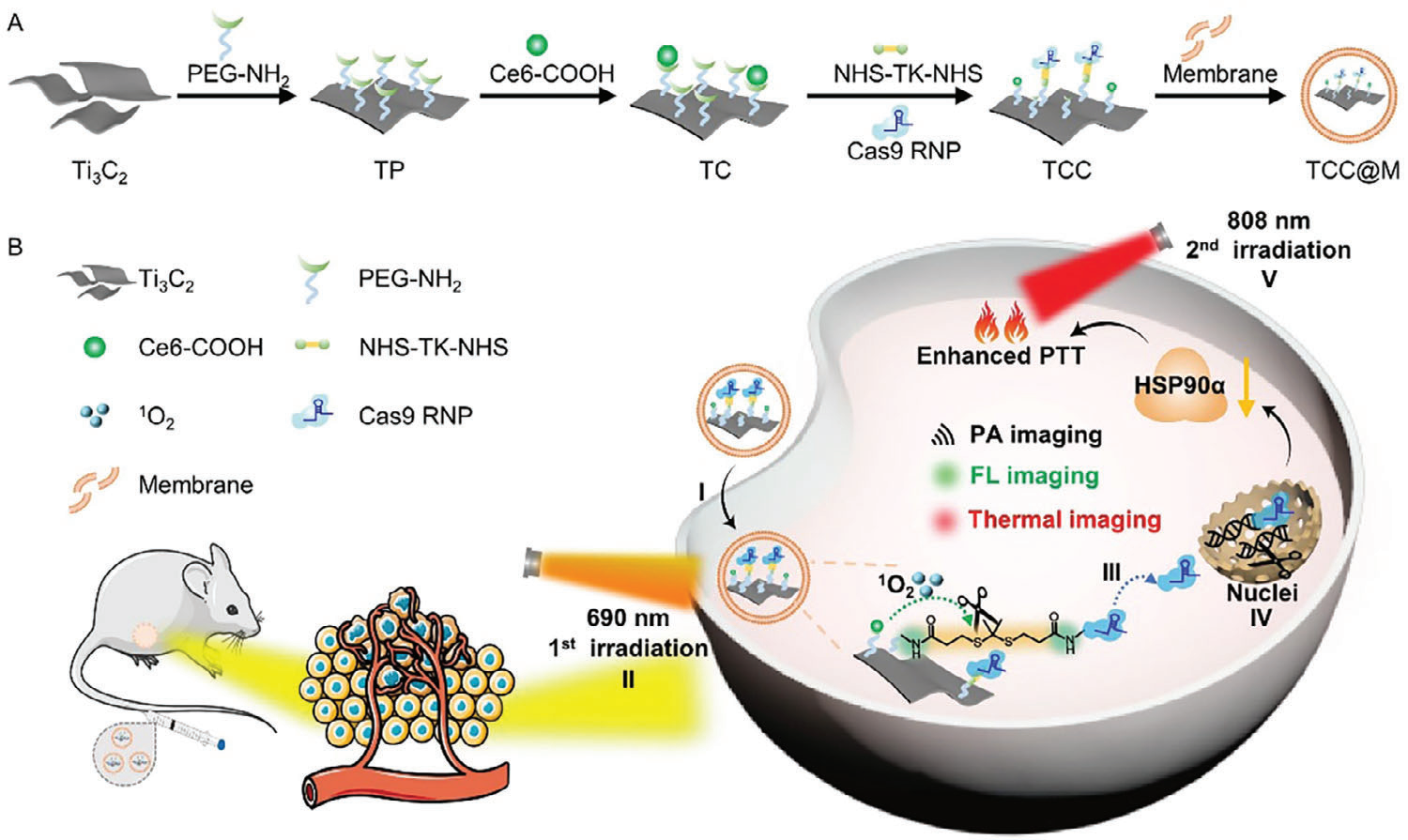
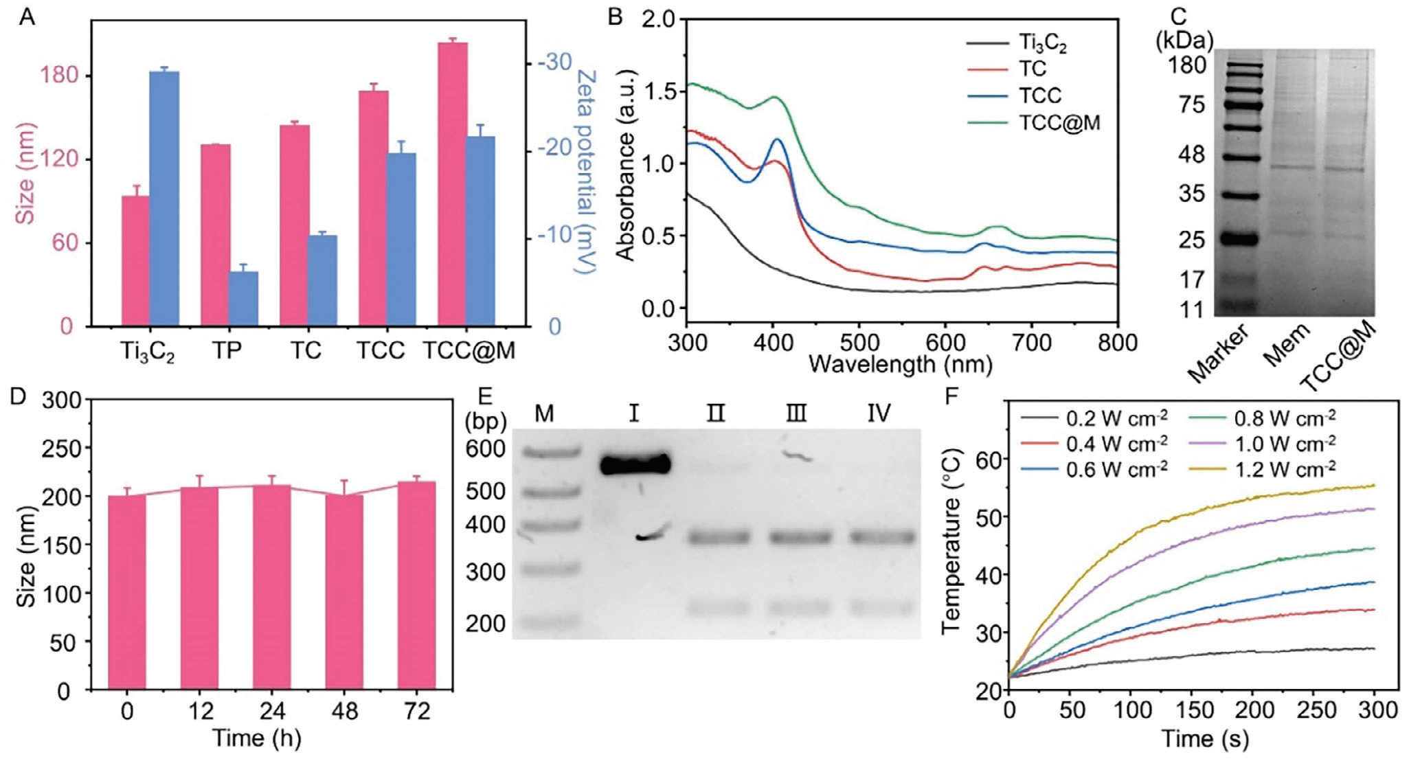
Experiment 1: Physicochemical Characterization of TCC@M
Core subjects: Physicochemical properties, stability and photothermal performance of Ti₃C₂, intermediate modified products and TCC@M.
Experimental logic: Gradient characterization verifies the successful step‑by‑step fabrication of TCC@M. The influences of cell‑membrane coating and light irradiation on Cas9 RNP activity are examined. Gradient experiments assess photothermal performance and stability.
Results: Figure 2 confirms successful step‑wise modification of TCC@M, retaining photodynamic and fluorescence‑imaging functions. The cell‑membrane coating is achieved with favorable aqueous stability. Cas9 RNP activity remains unaffected by membrane coating and light exposure, and excellent photothermal‑conversion performance is obtained.
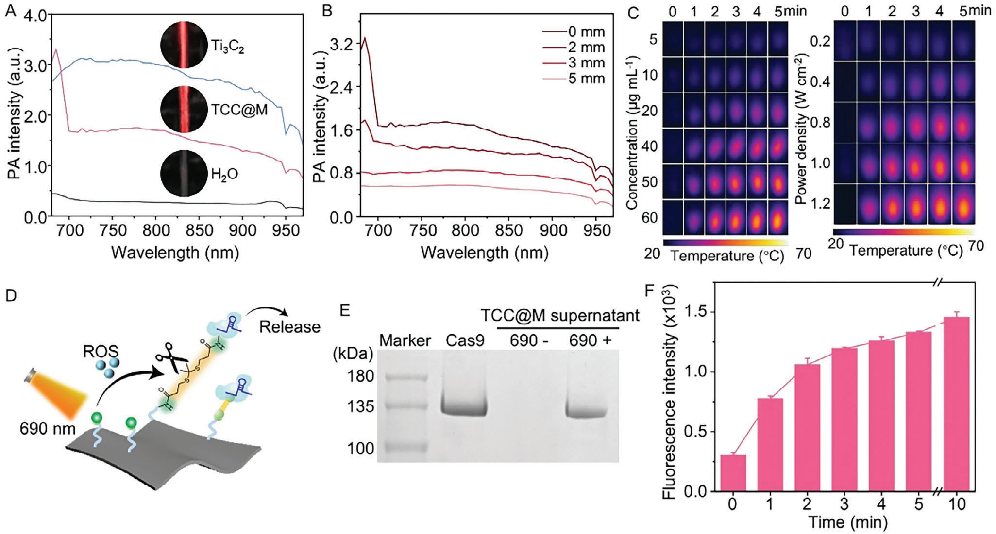
Experiment 2: Validation of Multi‑modal Imaging Performance and Light‑Triggered Cas9 RNP Release of TCC@M
Core subjects: Photoacoustic / thermal‑imaging performance of TCC@M, Cas9 RNP release efficiency and singlet‑oxygen generation capacity under 690 nm irradiation.
Experimental logic: *In‑vitro* imaging evaluates multi‑modal imaging capability. Electrophoresis and fluorescent labeling verify light‑triggered Cas9 RNP release. Probe‑based detection quantifies singlet‑oxygen yield.
Results: Figure 2 demonstrates TCC@M’s potential for photoacoustic imaging; 780 nm excitation is more suitable for *in‑vivo* photoacoustic imaging. Thermal imaging monitors temperature in real time. 690 nm irradiation effectively triggers Cas9 RNP release, and release efficiency rises with prolonged illumination time.
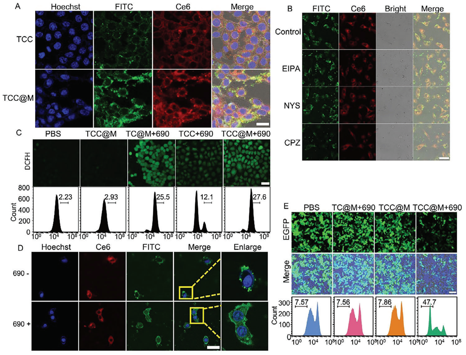
Experiment 3: Validation of Cellular Uptake, Endocytic Pathway and Nuclear Delivery of Cas9 RNP Mediated by TCC@M
Core subjects: Uptake efficiency and endocytic pathways of TCC@M in A549 cells; intracellular ROS production, lysosomal escape and nuclear delivery of Cas9 RNP upon light irradiation.
Experimental logic: Nexcope NCF confocal microscope is adopted for confocal imaging to compare uptake efficiency. Inhibitor assays identify endocytic pathways. Fluorescent probes detect ROS and lysosomal escape. Confocal imaging confirms nuclear localization of Cas9 RNP.
Results: Figure 4 shows higher cellular uptake of TCC@M attributed to homologous targeting. TCC@M enters cells mainly via clathrin‑dependent endocytosis. Light irradiation activates Ce6 to produce reactive oxygen species. Cas9 RNP successfully escapes lysosomes and achieves nuclear delivery with specific gene‑editing capability.
Experiment 4: Validation of TCC@M‑mediated HSP90α Gene Editing and Cellular Apoptosis
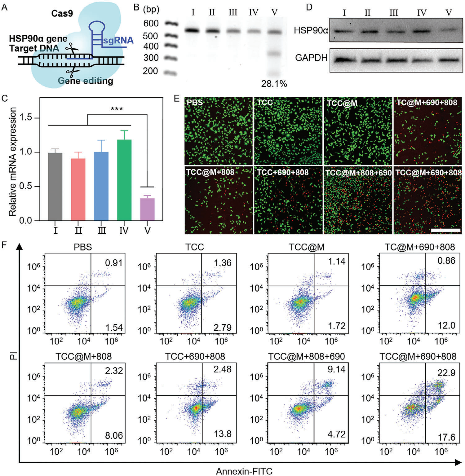
Core subjects: HSP90α gene‑cleavage efficiency, mRNA / protein expression in A549 cells; cell apoptosis and viability across different treatment groups.
Experimental logic: Restriction‑enzyme digestion verifies gene‑cleavage efficiency. qRT‑PCR and Western Blot detect gene / protein expression. Live‑dead staining and dual‑staining assays evaluate cell apoptosis and viability.
Results: Figure 5 proves that the TCC@M‑plus‑light group efficiently and specifically cleaves the HSP90α gene, markedly down‑regulating its mRNA and protein expression to thermally sensitize tumor cells. Sequential light irradiation significantly boosts tumor‑cell killing and apoptosis induction.
Experiment 5: *In‑Vivo* Multi‑modal Imaging of TCC@M and Optimization of Therapeutic Timing
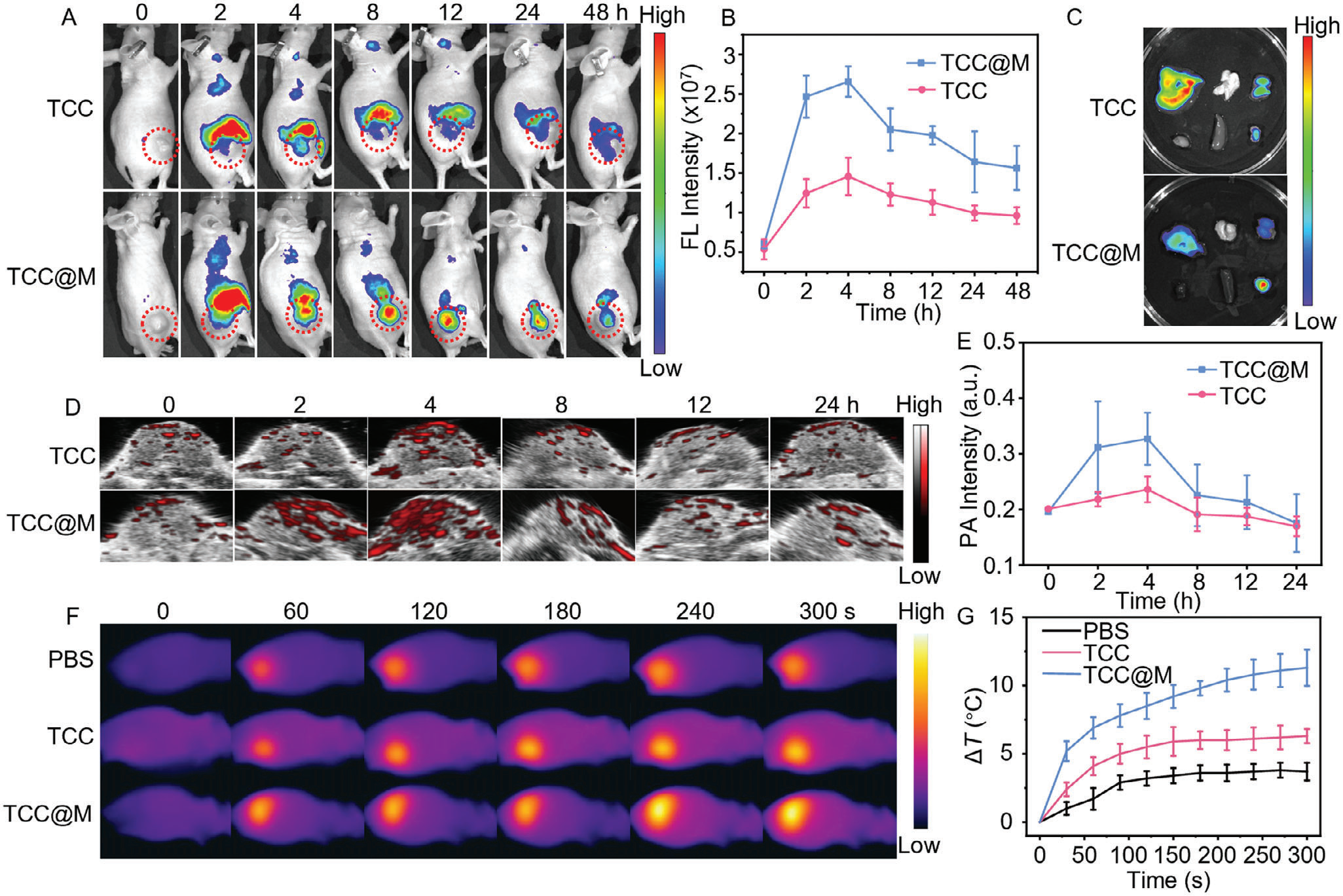
Core subjects: Fluorescence / photoacoustic / thermal‑imaging signals of TCC@M in A549 tumor‑bearing nude mice, tumor‑accumulation profile of the probe and optimal parameters for photothermal therapy.
Experimental logic: In‑vivo imaging dynamically monitors tumor‑site probe signals to determine peak‑accumulation time. Infrared thermal imaging tracks tumor‑temperature variation to optimize illumination parameters for photothermal therapy.
Results: Figure 6 reveals outstanding tumor‑targeting capability of TCC@M with peak accumulation at 4 h post‑injection. Photoacoustic signals under 780 nm excitation vary consistently with fluorescence signals. Illumination for 5 min at 4 h after injection represents the optimal timing and parameter for mild photothermal therapy.
Experiment 6: *In‑Vivo* Anti‑Tumor Efficacy and Histological Validation of TCC@M
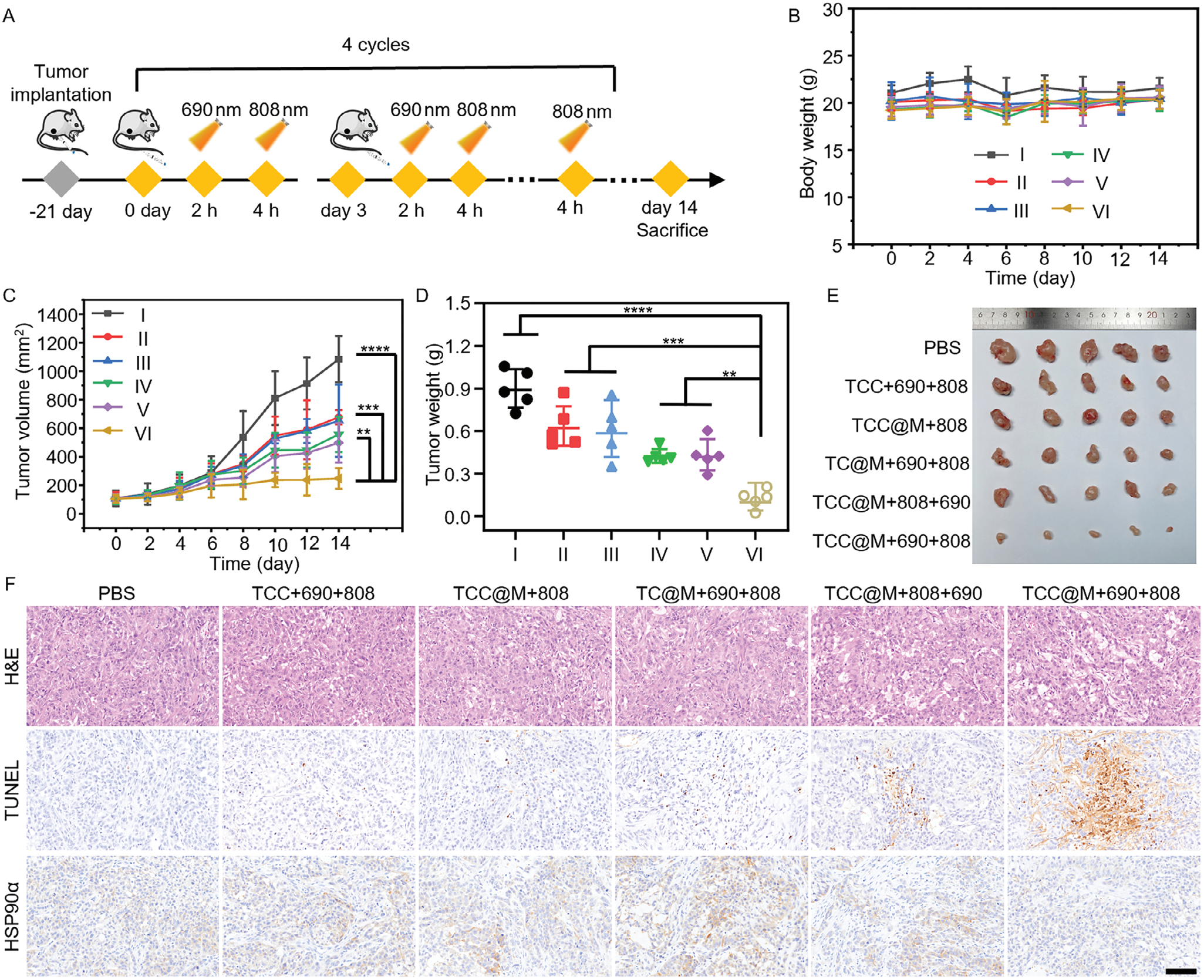
Core subjects: Tumor volume / weight, mouse body‑weight changes in A549 tumor‑bearing nude mice across groups; tumor histological features and major‑organ toxicity.
Experimental logic: Group‑controlled treatment assesses anti‑tumor efficacy and biosafety via relevant indexes. Histological staining identifies tumor necrosis and apoptosis and detects HSP90α expression. Organ staining evaluates systemic toxicity.
Results: Figure 7 demonstrates that the TCC@M sequential‑illumination group achieves far superior anti‑tumor effects versus control groups. All experimental groups exhibit good biocompatibility. This group shows the largest area of tumor necrosis and apoptosis plus drastically reduced HSP90α expression; only the non‑targeted group presents slight hepatic toxicity.
Conclusion
Relying on the Nexcope NCF confocal microscope for key cellular fluorescence‑imaging experiments, this study successfully constructed cancer‑cell‑membrane‑camouflaged Ti₃C₂ MXene nanoprobe TCC@M. The nanoprobe enables tumor‑targeted delivery with favorable photothermal performance and multi‑modal imaging capacities (fluorescence / photoacoustic / thermal). Triggered sequentially by two rounds of near‑infrared light, it first performs gene editing to thermally sensitize tumor cells, then induces enhanced photothermal therapy under mild temperatures for prominent tumor‑suppression outcomes. Multi‑modal imaging supports precise spatiotemporal control over cancer treatment.
Compared with conventional therapies, this technology overcomes pain points such as thermal resistance and poor synergistic effects. It features low off‑target effects, high targeting and excellent biocompatibility. It provides a novel precise‑theranostic platform for solid tumors including lung cancer. It can be further expanded to build multi‑target therapeutic systems. Its imaging techniques can be independently applied for early tumor diagnosis and intraoperative navigation, and supply a valuable experimental model for mechanism research in cancer therapy.