
Products
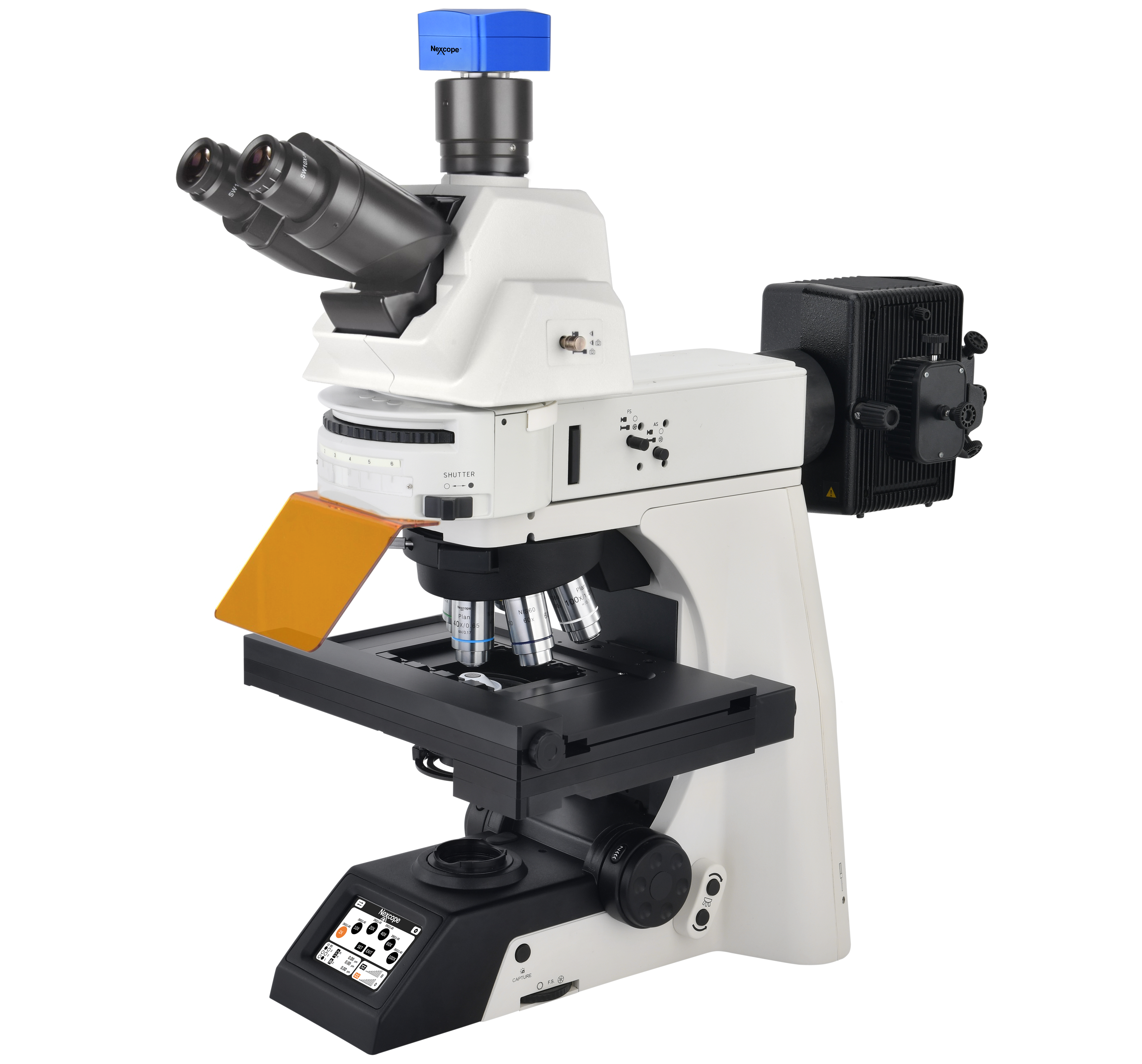
This advanced imaging platform delivers a large field‑of‑view for efficient observation of large‑scale samples. Integrated with multiple imaging technologies, it supports flexible optical‑path configurations and an optional Adaptive Focus System. Well‑suited for long‑term, high‑precision live‑cell observation, it is compatible with cutting‑edge modules such as confocal systems. Equipped with touch‑screen interaction, the system allows you to monitor sensors and control motorized components with one‑click scene switching to streamline workflows. Combined with powerful supporting software, it handles complex experiments with outstanding stability and expandability —‑ an ideal partner for modern life‑science research.
Stable, Highly‑Expandable Live‑Cell Imaging Platform
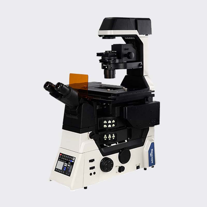
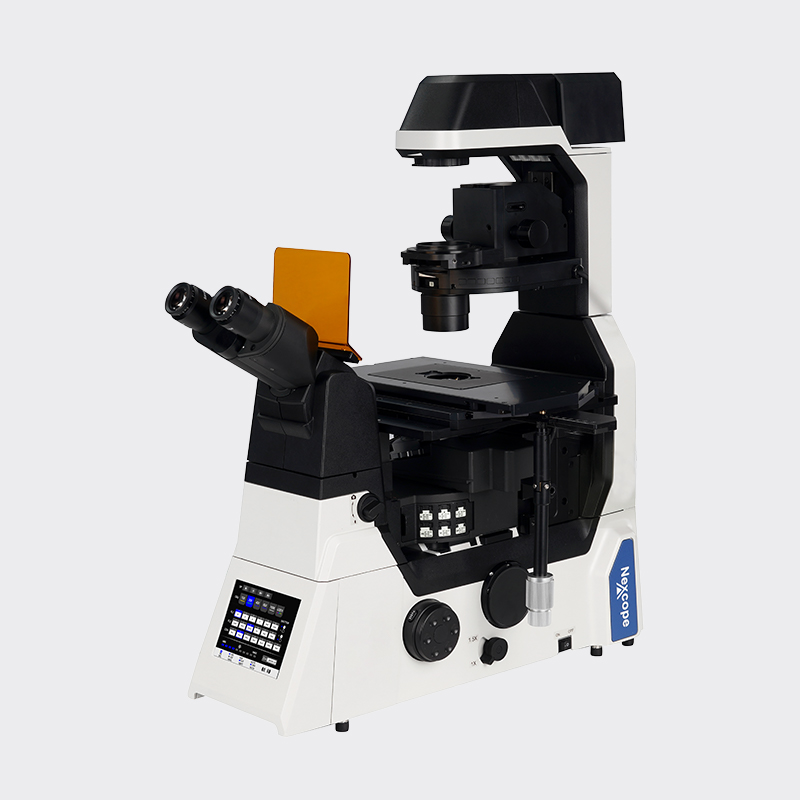
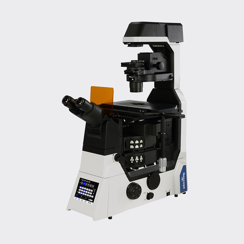
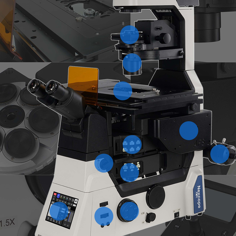
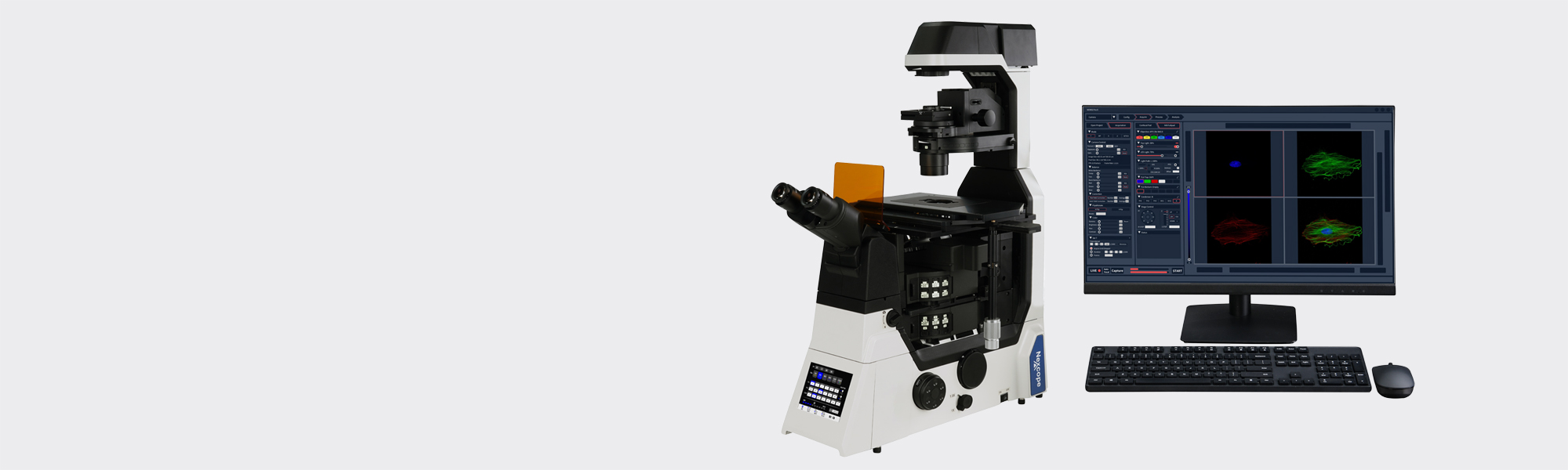
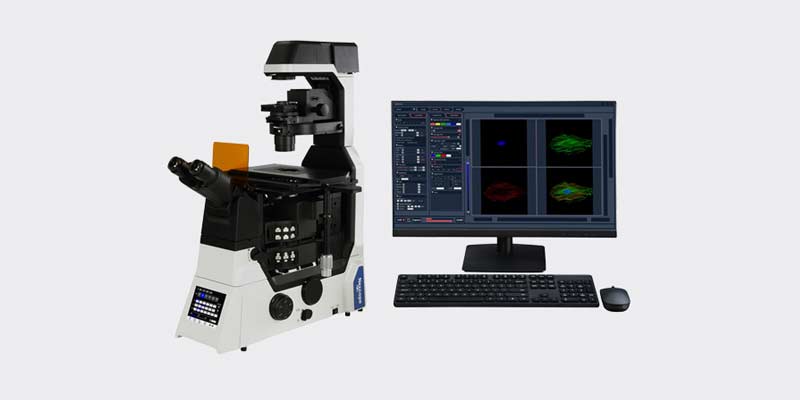
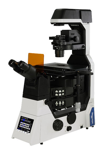
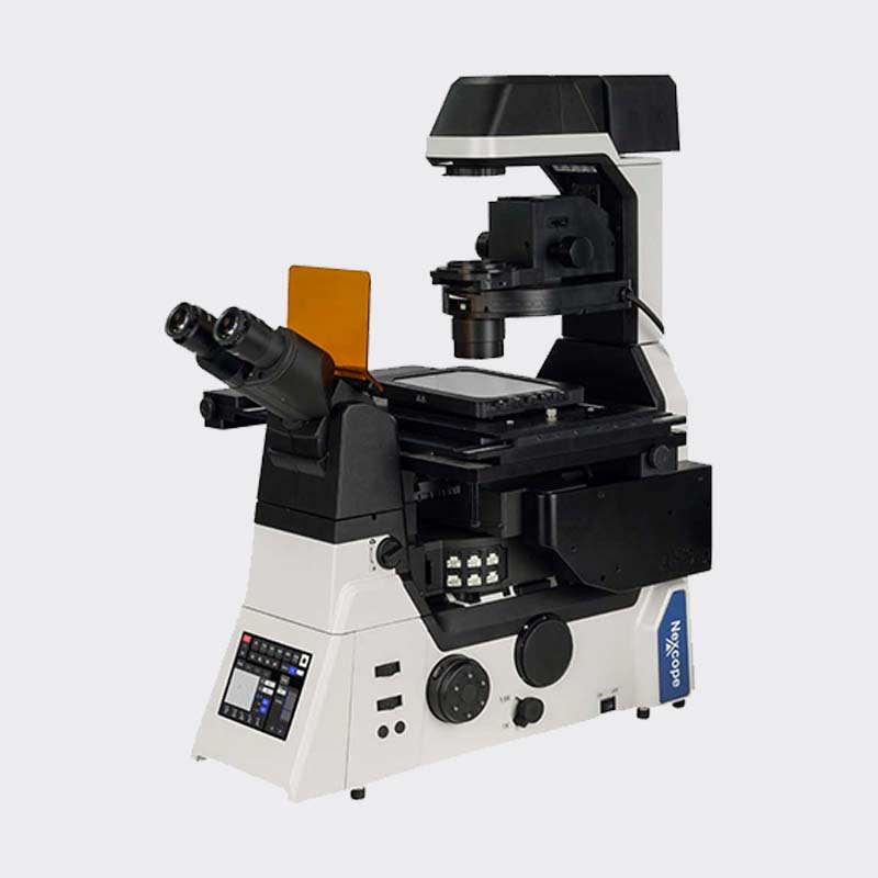
The NIB1000 microscope system is an advanced imaging platform for life‑science research, meeting analytical requirements for tissues, organs and living organisms. It includes two series: fully‑motorized AT and encoded M. Integrating bright‑field, fluorescence, DIC and phase‑contrast imaging technologies, it supports optional single‑ or dual‑layer optical paths. An optional Adaptive Focus System is available for long‑term, high‑precision live‑cell observation. It also reliably supports cutting‑edge imaging modalities such as confocal and super‑resolution microscopy. Equipped with touch‑screen interaction for easy one‑click scene switching, paired with professional image acquisition‑and‑analysis software, it delivers high stability and great expandability for complex experiments, ensuring reliable and reproducible data. It serves as an ideal tool for life‑science research.
NIB1000-AT
Single‑layer optical path

NIB1000-AT
Dual‑layer optical path

NIB1000-M
Single‑layer optical path

NIB1000-M
Dual‑layer optical path
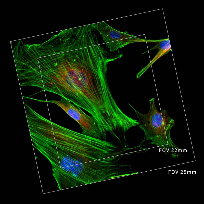
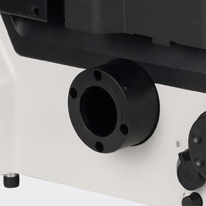
Unrivaled 25 mm Field‑of‑View
Research trends are shifting toward large‑scale, high‑throughput and intelligent solutions, creating growing demand for faster data acquisition and higher throughput capacity.
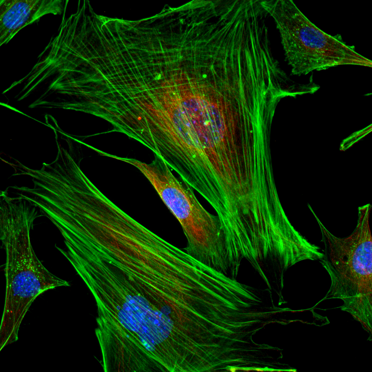
Whether for bright‑field or fluorescence imaging, the large‑area illuminator and wide‑field fluorescence accessories of the NIB1000 deliver uniform, bright microscopy images. Large‑format sensors and imaging interfaces fully unlock the potential of large‑area detectors, constituting an excellent microscopy imaging platform compatible with ongoing advances in camera technology.


Wide‑Field Illumination Module
Bright, large‑area transmitted illumination
Large‑aperture reflected‑fluorescence illuminator

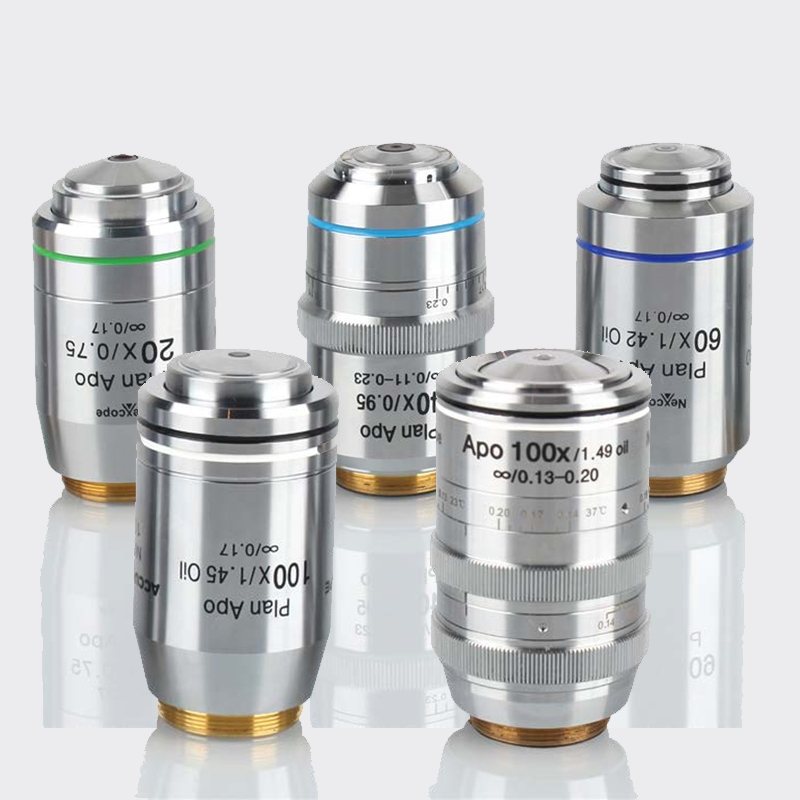
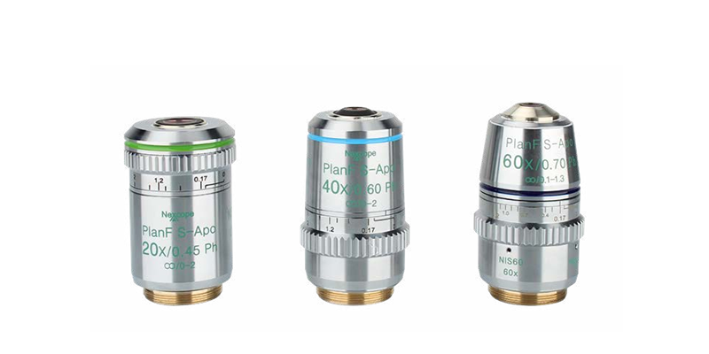
FOV25‑Capable Objectives
Premium objectives deliver outstanding field flatness for superior image quality. FOV25 objective field‑of‑view enables highly‑efficient data acquisition.

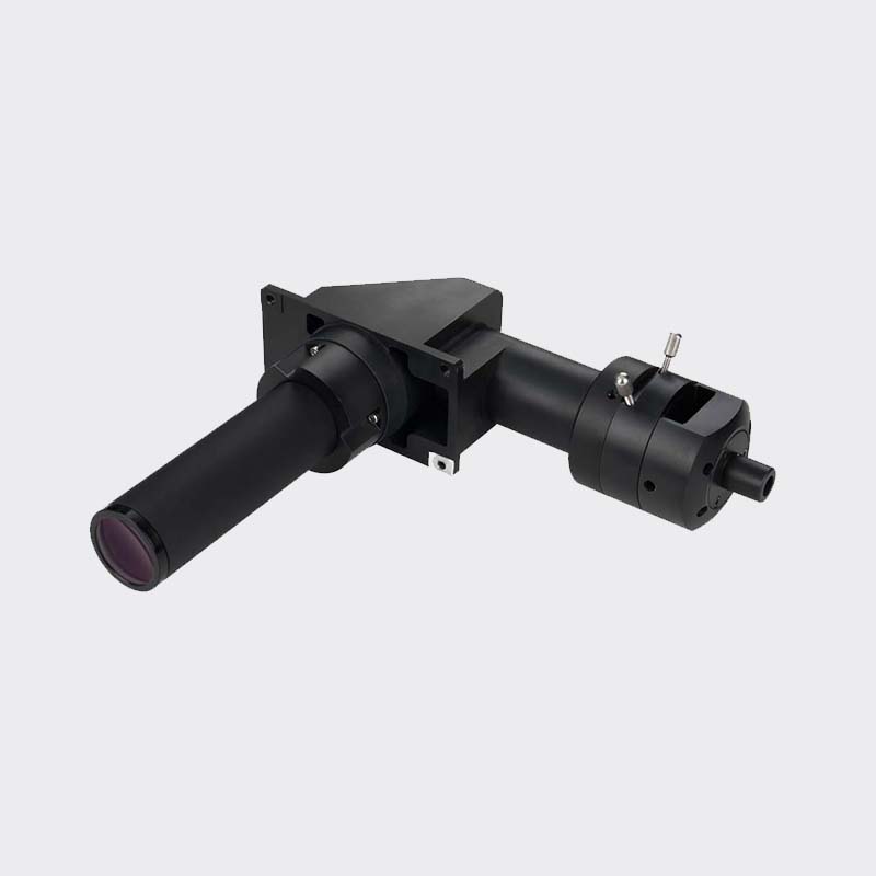
Large‑Aperture Observation Optics
Greatly improved light‑throughput performance. Delivers a broad 25 mm FOV for both bright‑field and fluorescence imaging with rich fine‑detail rendering.

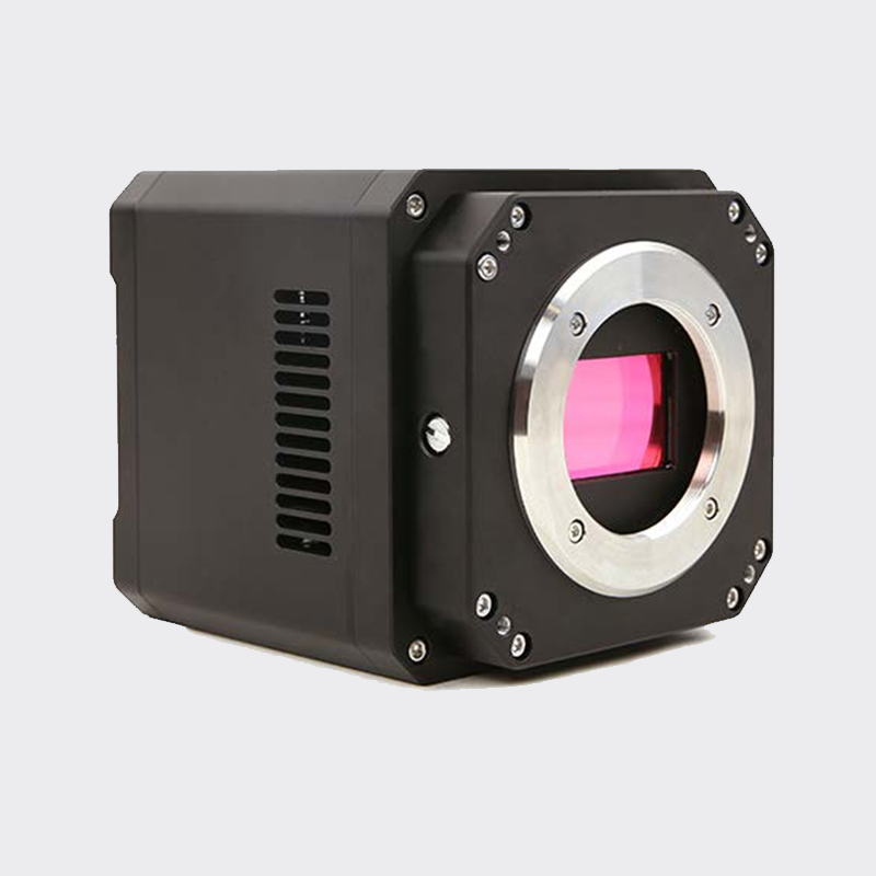
NEXCAM‑T24 High‑Sensitivity Monochrome Camera
36.0 × 23.9 mm CMOS sensor with 24‑megapixel resolution. Capable of ultra‑fast acquisition up to 114 fps, it provides ultra‑sharp, smooth digital imaging across the full field‑of‑view.

Live‑Cell Incubation System
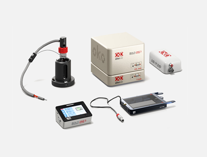
Specially designed for live‑cell imaging. Precisely regulates temperature across the microscope stage and inside culture dishes to maintain stable, uniform thermal conditions. The system also strictly controls humidity and CO₂ concentration to simulate physiological micro‑environments, providing stable and reliable incubation conditions for long‑duration live‑cell experiments.

Consistent, Reliable Imaging via AFS
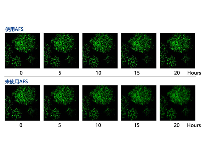
Featuring an independent focusing design to minimize mechanical influences on the Z‑axis. The new AFS (Adaptive Focus System) eliminates focus drift and maintains sharp focus under high‑magnification objectives as well as advanced modalities including super‑resolution, confocal and TIRF microscopy.

Premium LED‑Fluorescence Illumination System
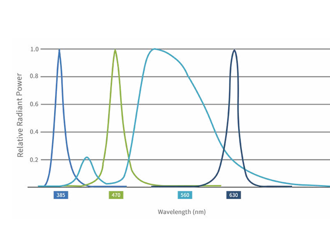
LED‑4 supports up to four‑channel LED illumination. It offers high compatibility with common fluorescent dyes, concentrated excitation energy and high brightness to satisfy routine experimental fluorescence‑imaging needs. Ready‑to‑use, long service life, no bulb replacement required. It reduces photobleaching and phototoxicity, making it highly suitable for live‑cell samples.
Intuitive, Ergonomic Controls
‑ Interactive operation: 5.7‑inch front‑panel touch‑screen display ‑ High‑speed motorized control: Efficient, convenient switching for objectives, filter cubes, stage and observation modules
The NIB1000 imaging system integrates advanced human‑machine interaction to deliver unprecedented fine‑control experience. Its intuitive, streamlined interface simplifies complex imaging workflows, enabling researchers to run sophisticated experiments efficiently and comfortably while reducing operator fatigue. It minimizes disturbance and damage to samples, capturing authentic, complete cellular dynamics to power life‑science research.
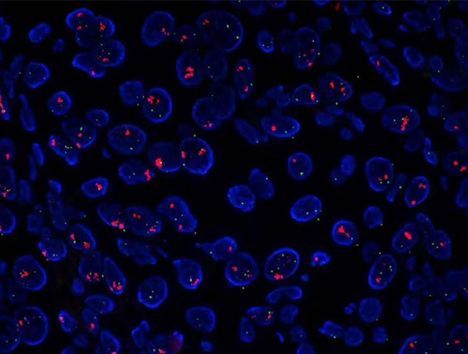

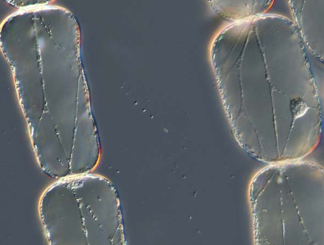
Phase‑contrast

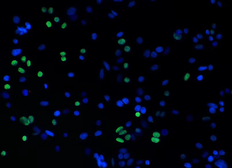
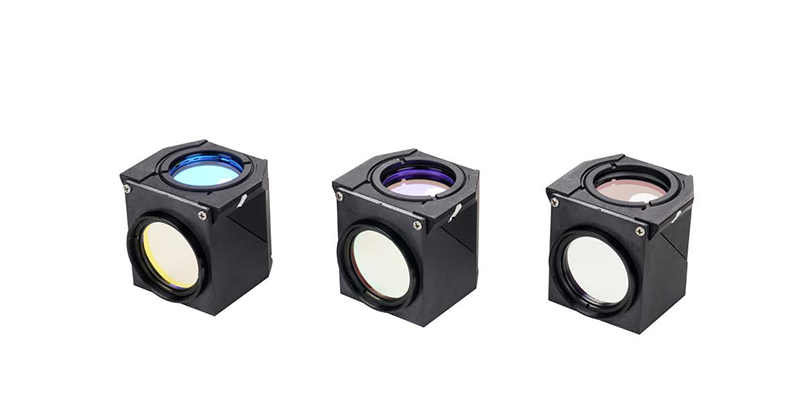
Fluorescence (filter cube set)


Nomis Pro X Microscopy‑Image‑Processing Software
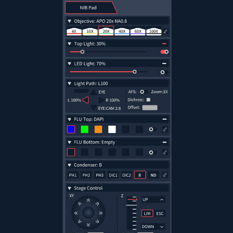
Nomis Pro X is Nexcope’s in‑house‑developed microscope software. It provides highly integrated control over core microscope functions and forms a high‑performance, user‑friendly all‑in‑one experimental solution.
For complex workflows or specialized research requirements, Nomis Pro X leverages outstanding integration and flexibility to deliver smooth operation. Researchers are freed from cumbersome microscope‑handling work and can focus on experiments and innovation.

Nomis Pro X Microscopy‑Image‑Processing Software
Nomis Pro X is Nexcope’s in‑house‑developed microscope software. It provides highly integrated control over core microscope functions and forms a high‑performance, user‑friendly all‑in‑one experimental solution.
For complex workflows or specialized research requirements, Nomis Pro X leverages outstanding integration and flexibility to deliver smooth operation. Researchers are freed from cumbersome microscope‑handling work and can focus on experiments and innovation.

Multi‑Dimensional Imaging
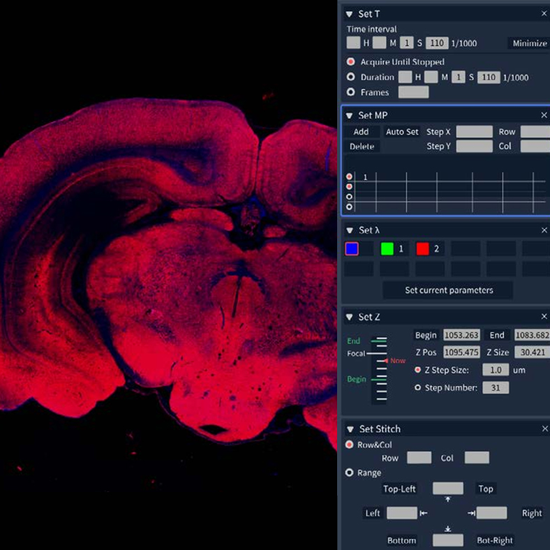
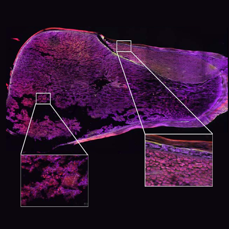
Stores custom observation presets for multi‑channel, multi‑position, time‑lapse and large‑tile‑stitching imaging workflows.

Deconvolution
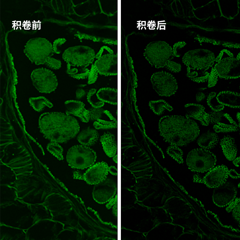
Performs deblurring for 2‑D images. Iterative deconvolution suppresses shot noise in images. 3‑D deconvolution is also available for multi‑dimensional datasets.

Multi‑Dimensional Image Visualization
Visualizes multi‑channel, time‑lapse, Z‑stack, XY‑position and image‑stitching information within a single window, improving experimental reproducibility.

High‑Speed Hardware Control
Software‑driven control of multiple motorized microscope components: objective turret, focus, condenser turret, fluorescence‑module switching and more.
Superb System Expandability
Can serve as the imaging platform for the NCF1000 laser‑scanning confocal microscope to generate low‑noise, high‑contrast, high‑quality confocal images.
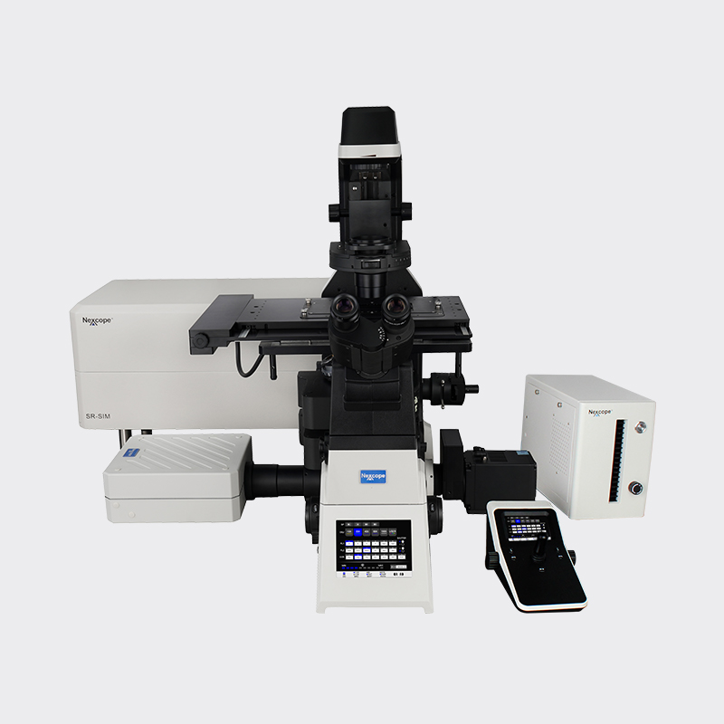
Compatible with the SIM super‑resolution system. The NSR1000 delivers live‑cell imaging at twice the resolution of conventional light microscopes.