
Products
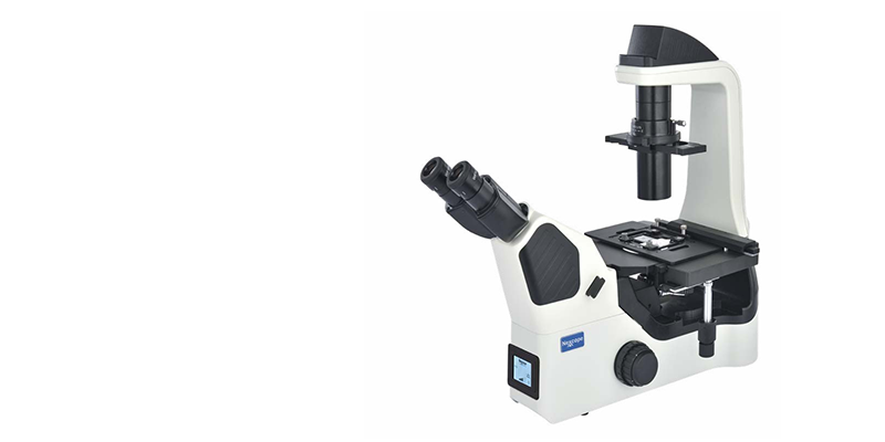
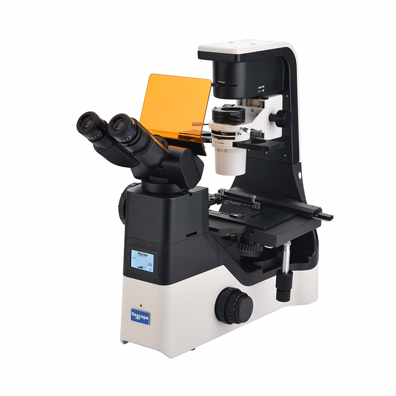
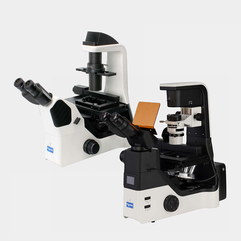
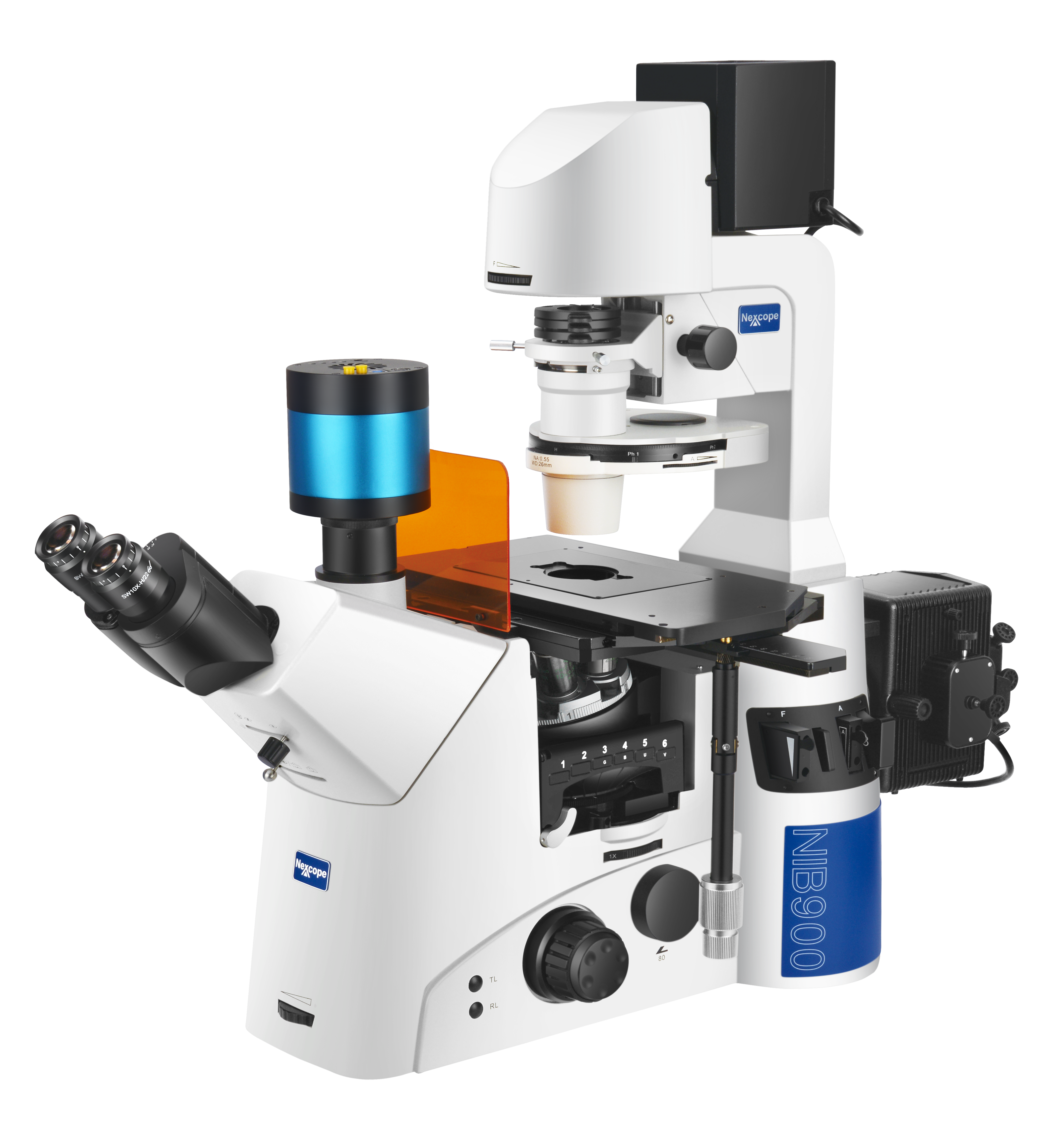
Refined from research‑grade microscopes, the NIB600‑Series is specially‑adapted for laboratory cell observation. Fitted with a long‑lifespan LED light source and infinity‑corrected optical system, it easily produces sharp, high‑contrast, wide‑field images. Its compact footprint and well‑arranged control buttons allow cell observation, sampling and sample processing inside a biosafety cabinet. Interchangeable filter sets provide greater flexibility for dye selection. High‑intensity, uniform‑brightness LED illumination supports high‑quality fluorescence imaging.
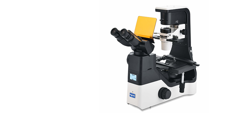
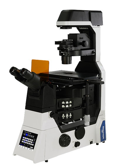
NIB650 / NIB660 Motorized Microscope

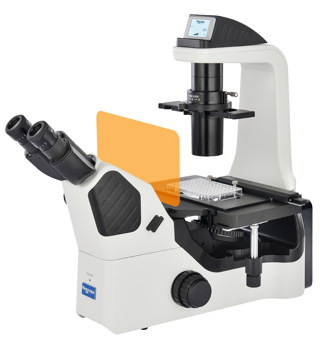
NIB620 / NIB630 Compact Cell‑Research Microscope

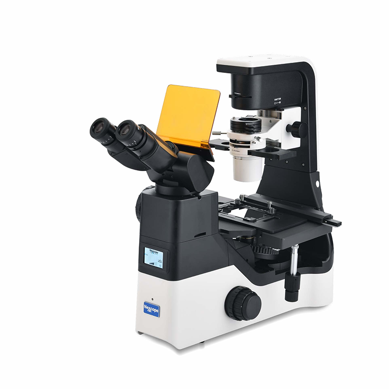
NIB610 / NIB620 Routine Cell‑Laboratory Microscope
Simple and Effective Imaging Solution Focused on Cell Observation
The NIB610/NIB620 cell‑culture microscopes are custom‑built for daily observation and research workflows in cell laboratories. Compared with conventional upright microscopes, they deliver superior operational flexibility and application adaptability. They support multiple cell‑observation modes including phase‑contrast, Hoffman Modulation Contrast and Relief 3D Contrast, enabling easy visualization of semi‑transparent live cells even within culture vessels. Featuring a compact footprint, it supports live‑cell observation, sampling and sample processing inside a confined clean‑bench environment. It builds a stable, efficient and continuous experimental workflow and serves as a reliable research assistant for cell‑observation applications.


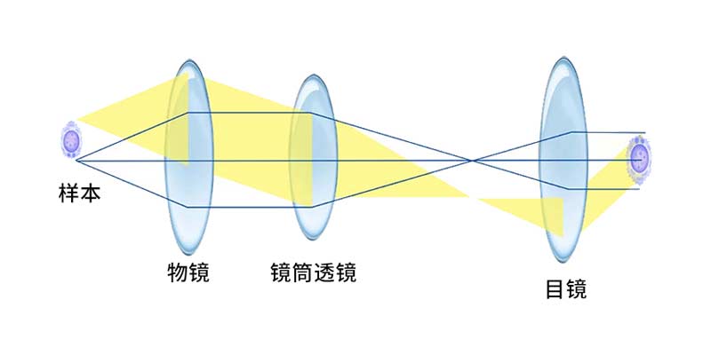
Infinity‑Corrected Optical System
The infinity‑corrected optical system minimizes aberrations and delivers uniform edge sharpness and color reproduction across the field‑of‑view. It fully unlocks resolution performance of high‑NA objectives and improves overall imaging resolution. Various optical modules can be flexibly added. A single microscope supports bright‑field, phase‑contrast, Hoffman Modulation Contrast, fluorescence and other observation modes.

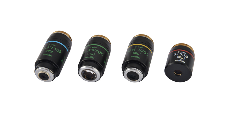
Specialized Objectives for Cell Observation
These objectives are purpose‑designed for cell observation and readily accommodate bright‑field, phase‑contrast, Hoffman Modulation Contrast, Relief 3D Contrast and other imaging requirements for diverse cell‑research scenarios. Optional semi‑apochromatic phase‑contrast objectives allow both phase‑contrast and fluorescence observation without objective replacement.
Hoffman Modulation Contrast
Relief 3D Contrast

Phase‑Contrast Observation
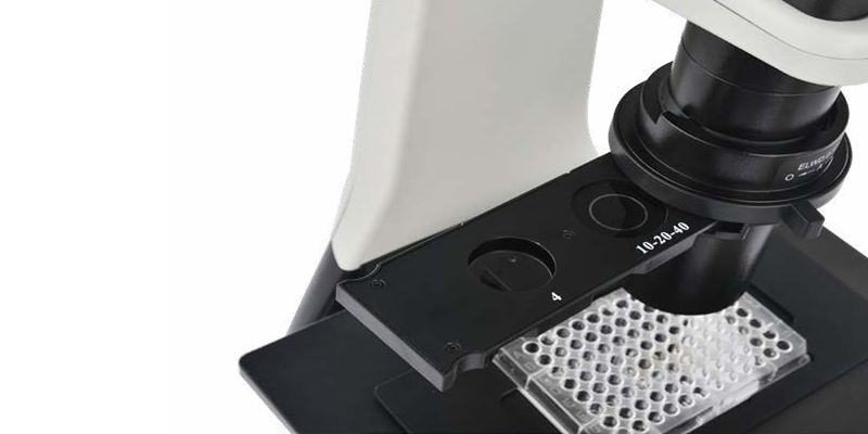
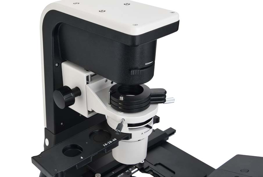
The NIB600 adopts integrated phase‑contrast sliders. Clear cell images at 4×, 10×, 20× and 40× can be obtained without replacing or readjusting phase rings.

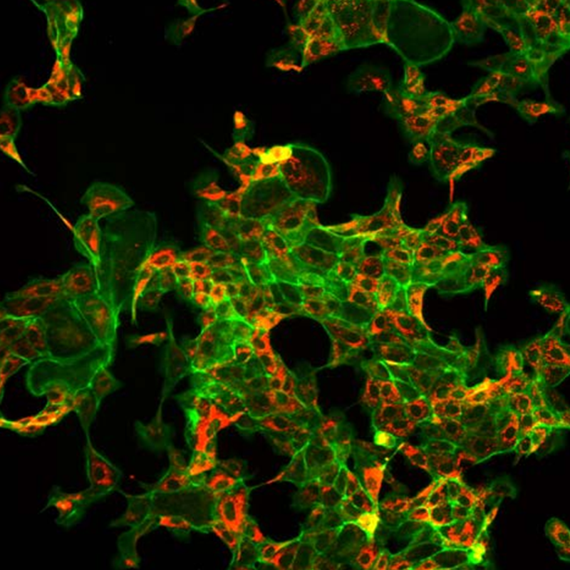
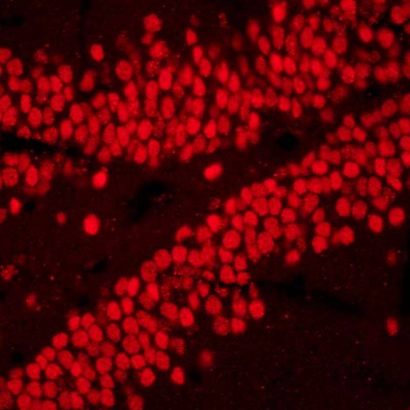
适用于多种荧光染料

Fly‑Eye Lens for
Uniform
Illumination

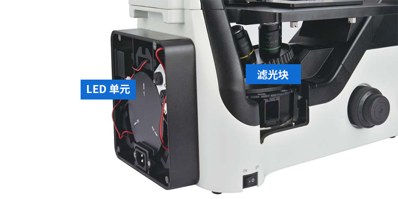
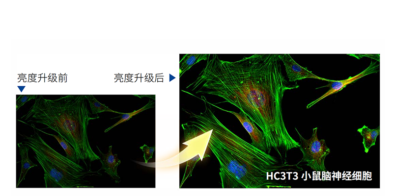
Newly‑Upgraded High‑Brightness LED Fluorescence Illumination

Encoded Objective Nosepiece
It memorizes illumination brightness for each objective. Light intensity is automatically adjusted upon objective switching, reducing visual fatigue and boosting work efficiency.

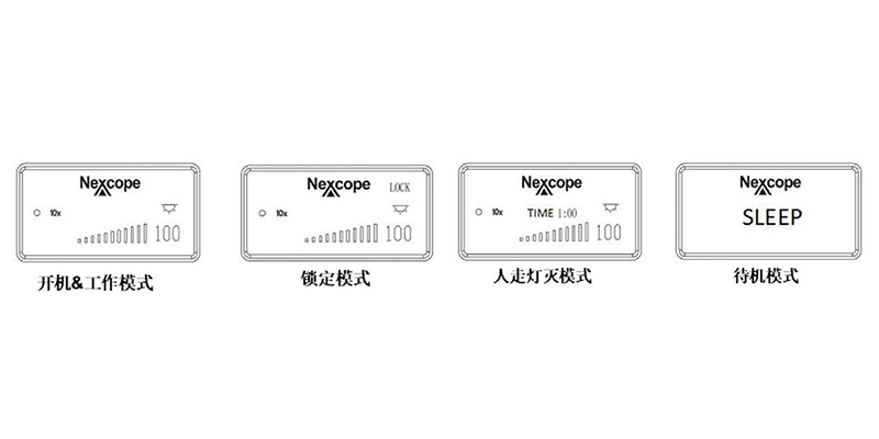
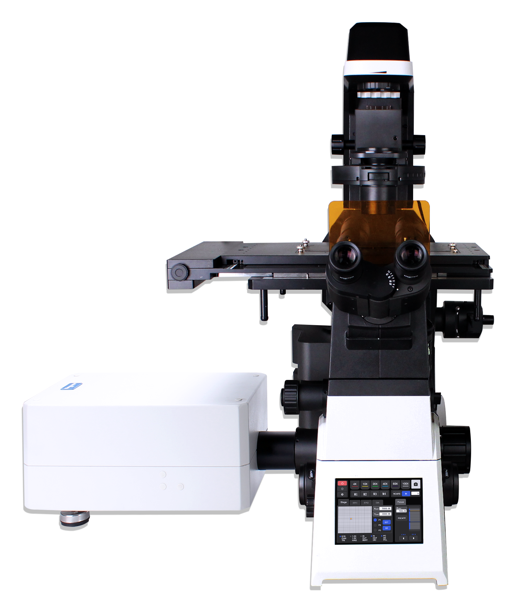
Microscope Status Display Screen
The front‑panel LCD screen displays microscope operational status including magnification, illumination intensity and standby status.
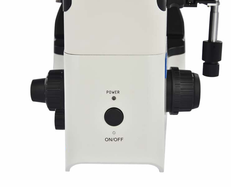
Well‑Laid‑Out Microscope Controls for Easy Operation
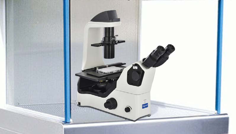
Compact, Stable and Small‑Footprint Body Suitable for Clean‑Bench Use
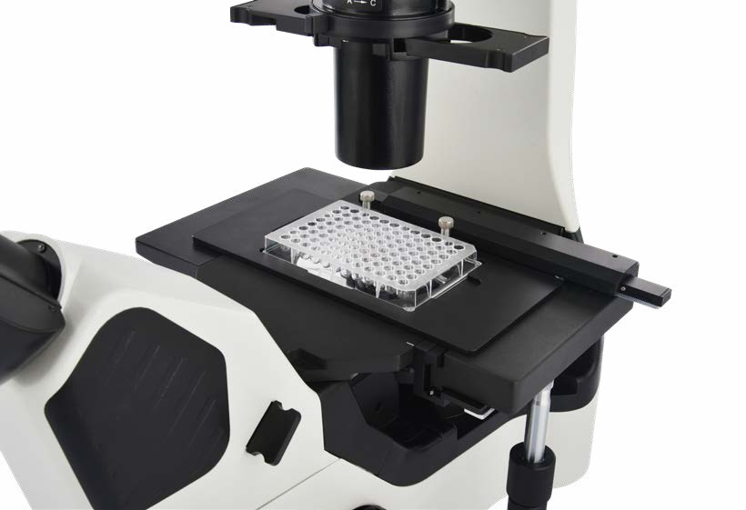
Multiple Stage Inserts for Various Cell‑Culture Vessels
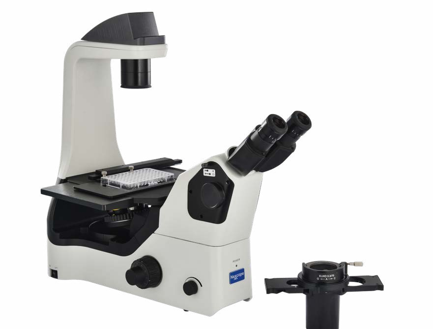
Removable Condenser
Deliver Highly‑Reproducible and Accurate Experimental Data for Cell‑Research Tasks
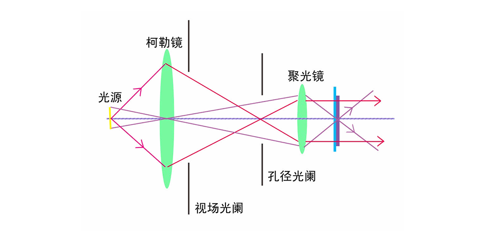
Within a compact footprint, the microscope features fully‑upgraded optics with Köhler illumination for markedly enhanced image sharpness and color fidelity. It precisely visualizes cell status and organelle morphology to support cell‑biology research. Thanks to the compact form‑factor of the NIB630, the unit fits easily inside laminar‑flow hoods for convenient operation, enabling delicate studies such as embryo micromanipulation. Ergonomic optimization eases visual and physical strain during long‑hour experiments, balancing comfort and experimental efficiency.


Illumination System with High Color‑Rendering Performance
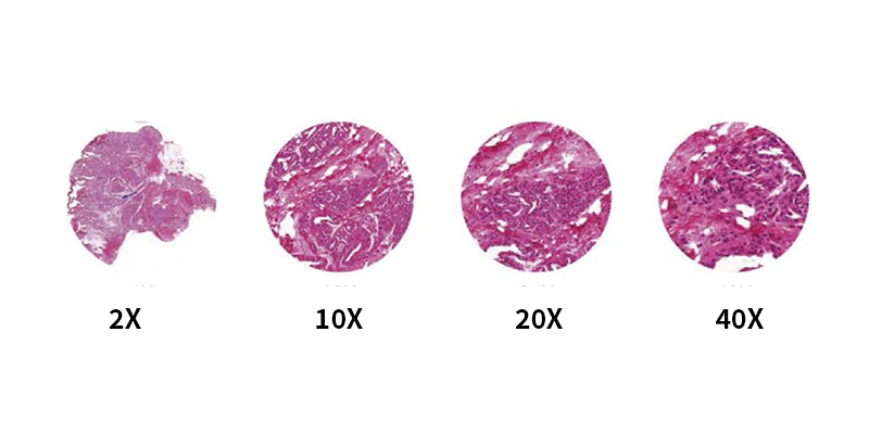
Equipped with Köhler illumination and daylight‑simulating LED light source, it delivers stable color temperature and uniform brightness. It faithfully reproduces true sample colors and distinguishes subtle color differences — a critical feature for pathological diagnosis.
Easily‑Adjustable Diaphragm Module
Easily‑accessible condenser‑aperture‑diaphragm controls. Adjust aperture‑diaphragm size to enhance imaging resolution, optimize image contrast and depth‑of‑field, extend the sharp viewing area and achieve optimal imaging results.

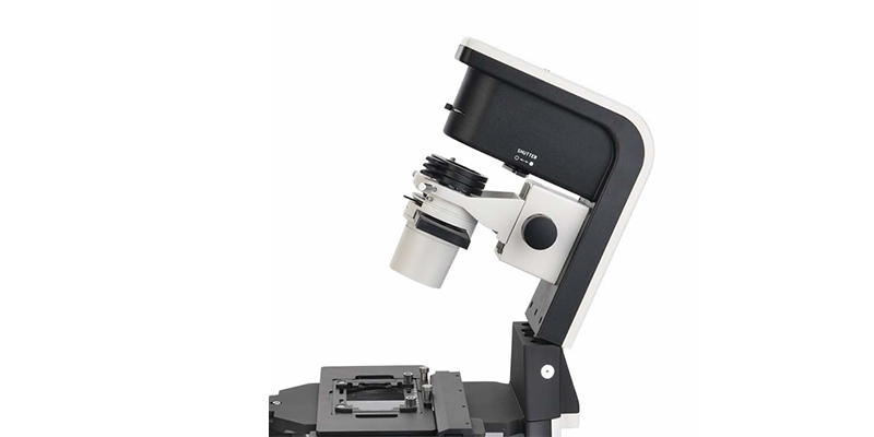
Tiltable Transmitted‑Illumination Column
Cell‑culture dishes are often large‑sized. The tiltable transmitted‑illumination column creates extra space for sample exchange, reducing movement distance and vibration during sample replacement.

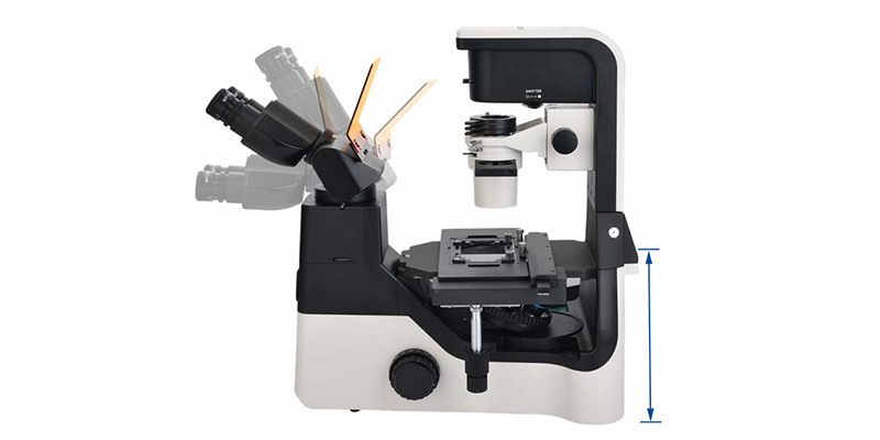
Comfortable Viewing Head
Ergonomic tiltable viewing head allows users to operate the microscope in a comfortable posture and minimizes muscle tension and discomfort caused by long‑duration work.

Highly‑Open Cell‑Experiment Platform — Build a Microscope Tailored for Your Team
‑ Motorized intelligent components to improve work efficiency
‑ Open architecture for customizing your dedicated microscope platform